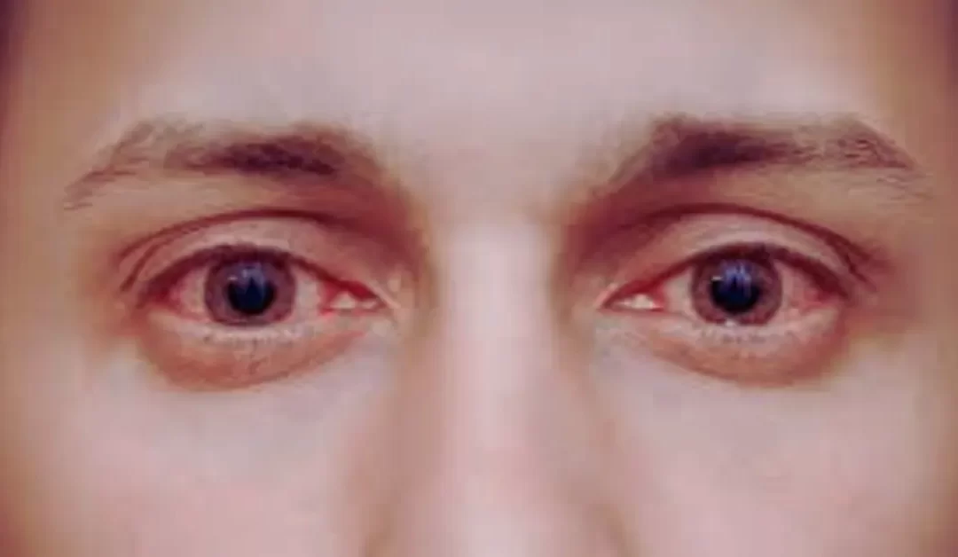
Vision begins with light, which reflects off objects and enters the eye if they’re within your field of vision. This intricate process involves various parts of the eye working together to transform light into the images you see.
The Tear Film and Cornea: Your Eye’s First Line of Focus
The first stop for light is the thin film of tears on the surface of the eye. This protective layer keeps the eye moist and clear. Behind it lies the cornea, the eye’s transparent front window. The cornea’s curved shape helps bend and focus incoming light toward the back of the eye.
The Aqueous Humor: Maintaining Balance
Behind the cornea is a liquid called the aqueous humor, which fills the space between the cornea and the lens. This clear fluid circulates within the front part of the eye, maintaining consistent pressure and nourishing nearby tissues.
The Pupil and Iris: Regulating Light
Light next passes through the pupil, a central opening within the iris, the colored part of your eye. The iris acts like a camera aperture, adjusting the size of the pupil to control how much light enters. In bright settings, the pupil contracts; in dim light, it expands to let in more light.
The Lens: Fine-Tuning Focus
After the pupil, light reaches the lens, which works like the lens of a camera to focus light precisely on the retina. The lens changes shape to accommodate objects at different distances, becoming thicker for nearby objects and thinner for distant ones.
The Vitreous Humor: A Jelly-Like Interior
Once light passes through the lens, it enters the center of the eyeball, a space filled with a clear, gel-like substance called the vitreous humor. This jelly maintains the eye’s shape and provides a medium for light to travel through.
Photoreceptors: Rods and Cones
Photoreceptors come in two types: rods and cones.
Rods are highly sensitive to low light and are responsible for peripheral and night vision.
Cones, on the other hand, function best in bright light and are essential for sharp central vision and color perception.
The Macula and Fovea: Precision Vision
At the retina’s center is the macula, a small, specialized area responsible for detailed vision. The very center of the macula, called the fovea, contains a dense concentration of cones, providing the sharpest focus and most vivid color detection.
Supporting Structures: RPE, Choroid, and Sclera
The retinal pigment epithelium (RPE) is a layer of dark tissue beneath the photoreceptors. It absorbs excess light to prevent scatter, ensuring clearer vision. The RPE also supports photoreceptors by transporting nutrients and removing waste.
The choroid is a vascular layer supplying oxygen and nutrients to the retina.
The sclera, the tough white outer layer of the eye, protects the delicate structures inside.
The Optic Nerve: Delivering Visual Signals
Signals generated by the photoreceptors travel along tiny nerve fibers within the retina. These fibers converge to form the optic nerve, which transmits visual information to the brain’s visual cortex.
The Brain: Interpreting the Image
The journey of light culminates in the brain, where the visual cortex processes the signals from the optic nerve. Here, the raw data is transformed into the coherent images we perceive as sight.
Seeing the World: A Complex Symphony
In summary, vision is the result of light reflecting off objects, entering the eye, being focused by the cornea and lens, converted into signals by the retina, and sent to the brain for interpretation. This intricate system works seamlessly to bring the world into focus.
Understanding how your eyes work highlights the complexity and wonder of human vision, an essential sense that connects us to the world around us.


